This study was reported according to the Strengthening The Reporting of Observational studies in Epidemiology (STROBE) guideline23. All methods were carried out in accordance with relevant guidelines and regulations. This study and its protocols were approved by the ethics committee of the Hamburg medical association. The patient data has been anonymized and therefore, according to the Hamburg medical association, informed consent was not needed (ethics vote WF-008/21).
We hypothesize that usage of fully threaded 3.5 mm titanium screws for IPSFC1/C2 is not suitable for establishing a spinal fusion between C1 and C2, in geriatric patients.
Patient population
Reviewing all posterior cervical operations at our institution in the time span from 2008–2012 we could identify 23 patients matching the inclusion criteria of an IPTSFC1/C2. The indication for surgical treatment was strictly determined in preoperative expert rounds. Indications were AAI after conservative therapy failure, unstable fractures, progressive osteolytic processes resulting in instability, or failure of other surgical therapy, such as anterior screw fixation. The respective indication for surgery for each patient is shown in Table 2. We performed a retrospective analysis of patients’ records and all available radiologic examinations in this regard.
Operative technique
Preoperatively, extensive diagnostics including cross-sectional imaging via computed tomography (CT) and magnetic resonance imaging (MRI) with visualization of the vertebral arteries is necessary to rule out contraindications such as the presence of a high riding vertebral artery (HRVA) or hyper kyphosis.
The operation was performed under general anesthesia. Fiberoptic awake intubation was performed due to the present cervical instability. In all cases, the C1/C2 Access System from DePuy Synthes (Oberdorf, Switzerland) and 3.5 mm, fully threaded, titanium alloy, cortical screws were used. A Mayfield clamp was applied, and the patient was positioned in a modified prone “concorde” position24. Fluoroscopic control of the anatomically correct position of C1 and C2 and in fracture cases verification of successful reduction, was applied. Sterile washing and draping according to the protocol followed. First, the correct entry point for the skin incision was located with the aid of a K-wire and marked (Fig. 3a). The K-wire was then placed percutaneously along the previously determined trajectory at the entry point for the screw, also under fluoroscopic control, on the C2 lamina, with help of a Jamshidi needle. The entry point for the screw is approximately 2 mm craniolateral to the medial edge of the caudal process of C2. After correct placement of the K-wire, the cortical bone of the lamina was punched with the K-wire to prevent slippage. Subsequently, a 2 cm skin incision was made, and the fascia was opened. A guide sleeve and then the tissue protection sleeve for the drill were passed over the K-wire to the entry point of the screw onto the lamina of C2 (Fig. 3b). From this point on, it was essential that the tissue protection sleeve always remained in firm bone contact and that the positioning at the entry point of the screw and the trajectory were not altered. If a dislocation occurred, removal of the sleeve, revisiting the screw entry point with the K-wire, and repeating the previous steps were necessary. If the sleeve remained securely on the lamina at the entry point, the screw channel was opened meticulously using a twist drill under fluoroscopic control. It was essential to keep the drill straight in the axis to avoid bending or breaking the drill and cutting osteoporotic bone. Therefore, an accurate fluoroscopic setting of the subsequent screw trajectory had to be set and constrained before the drill entered the bone. Once the drill entered the bone, no further correction could take place to avoid the above complications. Changes could only be achieved by complete repositioning. Once the screw channel had been opened successfully, the screw length was determined using the marking on the drill bit. In the case of particularly firm or sclerotic bone, this was followed by cutting a thread in the screw channel via the tissue protection sleeve, which remained permanently with firm bone contact at the entry point, also under fluoroscopic control. The 3.5 mm fully threaded screw was then inserted into the prepared screw channel with the previously determined length, using a self-retaining screwdriver, through the tissue protection sleeve under fluoroscopic control. Subsequently, the same procedure was followed for positioning the contralateral screw. Final fluoroscopic control of the screw position. After rinsing of the incisions, the wound was closed in layers with the reconstruction of the fascia, adapting subcutaneous suture and suture of the skin. Patients received a soft cervical orthosis for 6 weeks postoperatively. Post op cervical spine CTs were used to check for successful reduction and implant positioning.
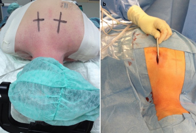
Intraoperative setting. After successful team time-out, a Mayfield clamp was applied, and the patient was positioned in a modified prone position, the so-called Concorde position. (a) First, the correct entry point for the skin incision is located with the aid of a K-wire and marked, usually at the level of TH 3/4 approximately 3 cm to the midline. (b) Subsequently, a 2 cm skin incision is made, and the fascia is opened. The drill guide is now advanced thrue the para spinal muscles and is placed on the lateral mass of C2. This is followed by fluoroscopically controlled drilling with constant monitoring for bone contact with the drill. When the drilling depth is sufficient according to the x-ray, the instrument provides the possibility of measuring the screw length. The screw can then be inserted through the guide instrument, again under fluoroscopic control.
Clinical and radiological evaluation
Preoperative and all postoperative imaging, consisting of conventional radiography (CR) and CT, were evaluated by the first and last author independently. Questionable cases were reevaluated by the spine surgeon M.S. Postoperative imaging was evaluated for implant positioning, possible implant loosening and the presence of spinal fusion. As reported by Kaminski et al.4 we evaluated the implant positioning according to the criteria published by Madawi et al.25. The implants position was considered appropriate if the screws were bridging the atlantoaxial joints on both sides, passing the lateral mass of C2 and C1 on both sides and did not protrude more than 5 mm beyond the anterior arch of C1 (Fig. 4a,b). Screws that did not meet all criteria were considered to be malpositioned. If there was a lightening halo around the screws on the follow up CR or CT scans, we assumed implant loosening. The presence of spinal fusion was assumed if there was radiographically clear bony bridging between C1 and C2. The following parameters were collected to evaluate the clinical outcome of the patients. Postoperative hospitalization time and time in the ICU were recorded. Furthermore, we evaluated the patients’ pain symptoms pre- and postoperatively by means of a 10-point visual analogue scale (VAS). Peripheral neurological assessment of the patients was performed pre- and postoperatively as well as at last follow-up by means of the Frankel scale26.

Correct screw position. This figure shows a typical example of a correct material position after isolated percutaneous transarticular screw fixation of C1 and C2 (IPTSFC1/C2). Conventional X-ray. (a) Anterioposterior plane. The screws inserted bridge the atlantoaxial joints on both sides and pass thrue the lateral mass of C1 and C2 on both sides. (b) Sagittal plane. The screws do not protrude more than 5 mm beyond the anterior arch of C1. If all these criteria match, the position of the screw is considered to be correct.
Statistical analyses
For statistical analysis, the software SigmaPlot 13 of Systat Software Inc., San Jose, CA, USA was used. The analysis of the patient data was descriptive. Continuous variables are expressed as mean ± standard deviation. Exceptions are found in the values of follow-up time. These values are given in median, range, and quartiles (Q1/Q3). Categorial variables are expressed as number and/or percentage. The Shapiro–Wilk test was used to test normal distribution. To evaluate the statistically significant differences between the preoperative and follow-up measurement time point, t-test for dependent samples in the case of a normal distribution or the Wilcoxon rank sum test in the case of a non-normal distribution was used. The significance level was p < 0.05.
Ethics declaration
This study was reported according to the Strengthening The Reporting of Observational studies in Epidemiology (STROBE) guideline23. All methods were carried out in accordance with relevant guidelines and regulations. This study and its protocols were approved by the ethics committee of the Hamburg medical association. The patient data has been anonymized and therefore, according to the Hamburg medical association, informed consent was not needed (ethics vote WF-008/21).